



 Save Product

Product Details
Application
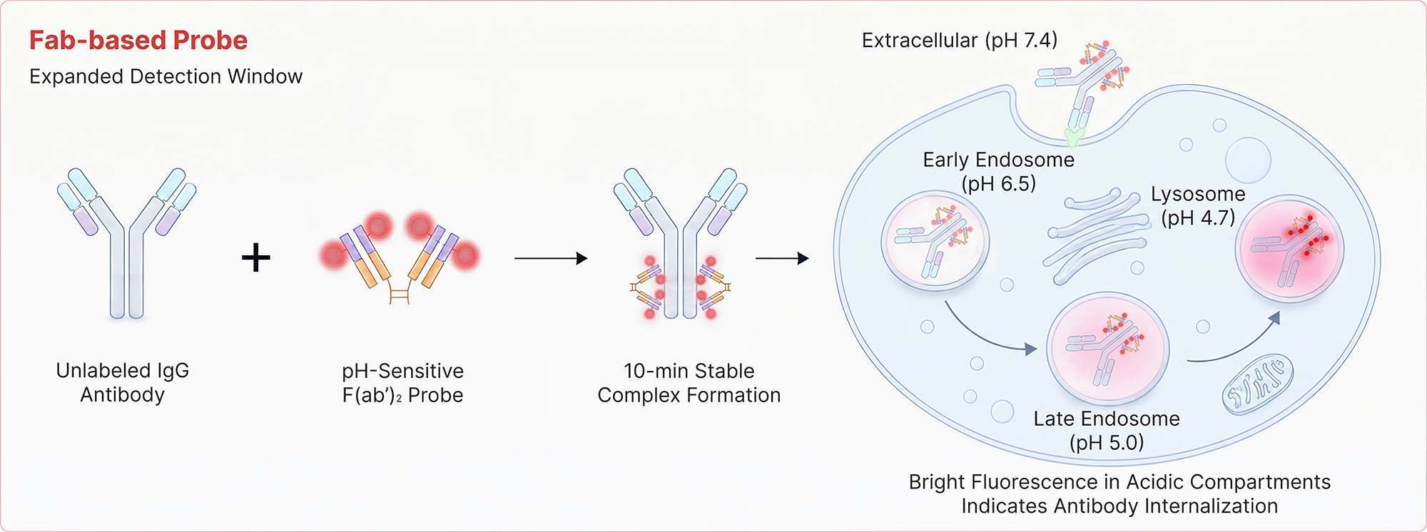
Antibody Internalization Detection Reagent (Cat. No. IGG‑PZF2001) utilizes a goat polyclonal anti‑human IgG Fc antibody conjugated to a pH‑sensitive fluorescent dye, specially engineered for acid‑triggered signal amplification. This reagent specifically labels the Fc region of human IgG antibodies (IgG1, IgG2, IgG3, and IgG4) to form a stable complex, enabling real‑time monitoring of antibody endocytosis in live cells. The pH‑sensitive dye not only significantly enhances the fluorescence signal in an acidic environment but also minimizes background noise.
Features
- High signal-to-noise ratio - Strong fluorescence with minimal background
- Rapid fluorescent labeling - Complete in 10 minutes
- pH sensitive - Bright emission in acidic milieu
- Fc-Specific targeting - Preserves Fab antigen-binding integrity
Specificity
Specifically recognizes the human antibody Fc portion.
Detection Wavelength
Excitation Wavelength: 643 nm
Emission Wavelength: 660 nmStorage
For long term storage, the product should be stored at lyophilized state at -20°C or lower.
Please protect from light and avoid repeated freeze-thaw cycles.
This product is stable after storage at:
- -20°C to -70°C for 24 months in lyophilized state;
- -20°C to -70°C for 12 months after reconstitution.
Assay Principles
ACRO Quality Management System
Customers Also Viewed
Performance Data
Bioactivity-Flow Cytometry

Anti-CD20 Abs and Human IgG1 isotype control were labeled with Antibody Internalization Detection Reagent (Cat. No. IGG-PZF2001). Raji cells were treated with Anti-CD20 Abs-Internalization Detection Reagent conjugate and Isotype control-Internalization Detection Reagent conjugate separately for 2 hours, then analysis by Flow cytometric. APC signal was used to evaluate the activity (QC tested).
Protocol
Anti-Her2 Abs and Human IgG1 isotype control were labeled with Antibody Internalization Detection Reagent (Cat. No. IGG-PZF2001). SK-BR-3 cells were treated with Anti-Her2 Abs-Internalization Detection Reagent conjugate and Isotype control-Internalization Detection Reagent conjugate separately for 2 hours, then analysis by Flow cytometric. APC signal was used to evaluate the activity (Routinely tested).
Protocol
Trontinemab and Human IgG1 isotype control were labeled with Antibody Internalization Detection Reagent (Cat. No. IGG-PZF2001). HEK293/Human Transferrin R Stable Cell Line were treated with Trontinemab-Internalization Detection Reagent conjugate and Isotype control-Internalization Detection Reagent conjugate separately for 4 hours, then analyzed by Flow cytometric. APC signal was used to evaluate the activity (Routinely tested).
Protocol
FACS Data

In the assessment of ADC efficacy using the Antibody Internalization Detection Reagent (ACROBIOsystems, Cat. No. IGG-PZF2001) with HER2-positive SK-BR-3 cells and HER2-negative MFI-MDA-MB-468 as target cells; (A) Prolonged co-incubation of HER2-targeting antibodies with HER2-positive SK-BR-3 cells enhanced antibody internalization. (B) whereas no internalization was observed in HER2-negative MFI-MDA-MB-468 cells.
Protocol
In the assessment of ADC efficacy using the Antibody Internalization Detection Reagent (ACROBIOsystems, Cat. No. IGG-PZF2001) with HER2-positive SK-BR-3 cells as target cells; (C) The trastuzumab-based ADC effectively internalizes relative to its naked antibody counterpart at concentrations ≥0.1 μg/ml, demonstrating dose-dependent enhancement with comparable efficacy between the two. (D) Disitamab-based ADC effectively internalizes relative to its naked antibody counterpart at concentrations ≥0.1 μg/ml, demonstrating dose-dependent enhancement, with no statistically significant difference observed between the two.
Protocol
Fluorescence Imaging

SK-BR-3 cells were treated with CellLights Lysosome GFP (green) for 16 hours followed by treatment with Anti-Her2 Abs-Internalization Detection Reagent conjugate and IgG1 Isotype-Internalization Detection Reagent conjugate separately for 16 hours (red), then stained with NucBlue Live ReadyProbes(blue) for 20 minutes and imaged on the EVOS M7000. A. Antibody Internalization Detection Reagent (Cat.No.IGG-PZF2001). B. IgG1 Isotype-Internalization Detection Reagent conjugate. C. Anti-Her2 Abs-Internalization Detection Reagent conjugate. D. Anti-Her2 Abs-Internalization Detection Reagent conjugate(Z-stacking).
Customer Reviews Writing Reviews

FAQ
- product
Can lyophilized proteins remain stable during room-temperature shipping or temporary room-temperature exposure?
In general, lyophilized proteins remain stable during room-temperature shipping or temporary exposure to room-temperature conditions.
Many ACROBiosystems recombinant proteins are supplied in lyophilized (freeze-dried) form. The lyophilization process effectively removes moisture from the product, helping reduce degradation-related reactions and improve protein stability during transportation and storage.
To evaluate the stability of lyophilized proteins under normal temperature conditions, ACROBiosystems conducted a stability study on 11 representative lyophilized protein products at 37°C. The study demonstrated that these proteins maintained good quality stability after storage at 37°C for approximately 20 days. Based on these validation results, temporary room-temperature exposure during shipping or handling is generally not expected to have a significant impact on product quality or downstream application performance.
To ensure optimal long-term stability, products should be stored according to the storage conditions specified in the product Certificate of Analysis (COA) upon receipt.
Supporting Document
Recent Advances
 Contact Us
メールアドレス
order.jp@acrobiosystems.com
お問い合わせ
グローバル:+1 800-810-0816(Toll Free)
 Drug Candidate Licensing