Development and validation of bioanalytical assays for the quantification of ADC
 Publication Date:2026-06-17
 Page Views:336
Introduction
Antibody-drug conjugates (ADCs) represent a novel class of targeted cancer therapeutics that strategically combine the exquisite specificity of monoclonal antibodies (mAbs) with the potent cytotoxic payloads.1 By precisely delivering these cytotoxic payload to malignant cells, ADCs aim to significantly enhance the therapeutic window-maximizing efficacy while minimizing systemic toxicity. As the ADC field continues to expand with more than 20 FDA-approved products and numerous candidates in clinical development, comprehensive characterization of their pharmacokinetic (PK) profiles has become fundamental for evidence-based dosing strategies, safety assessments, and efficacy optimization.2
Unlike conventional therapeutics, ADCs present unique analytical challenges stemming from their structural and functional complexity. Their heterogeneous drug-to-antibody ratios (DARs), diverse linker chemistries, payload properties, and dynamic in vivo biotransformation pathways necessitate sophisticated bioanalytical methods.3 Accurate quantification of multiple ADC-related analytes is critical not only for PK assessment but also for establishing meaningful pharmacodynamic (PD) relationships, evaluating toxico-kinetic profiles, and predicting therapeutic outcomes.3
Comprehensive PK characterization of ADCs typically requires quantification of four distinct analytes:
- Total antibody (Tab): Encompasses all antibody species regardless of conjugation status (DAR ≥ 0).4
- Conjugated antibody: Quantifies antibody species with at least one attached payload (DAR ≥ 1).4
- Conjugated drug: Measures the payload amount that remains covalently linked to the antibody.4
- Free drug: Quantifies the unbound cytotoxic payload in circulation.4
These complementary analytes collectively illuminate different aspects of ADC. Total antibody measurements reveal whether conjugation fundamentally alters the antibody's inherent PK properties. Conjugated antibody and drug concentrations provide insights into in vivo stability and DAR dynamics over time. Free drug levels serve as critical safety indicators by tracking potential systemic exposure to the off-target toxicity.4
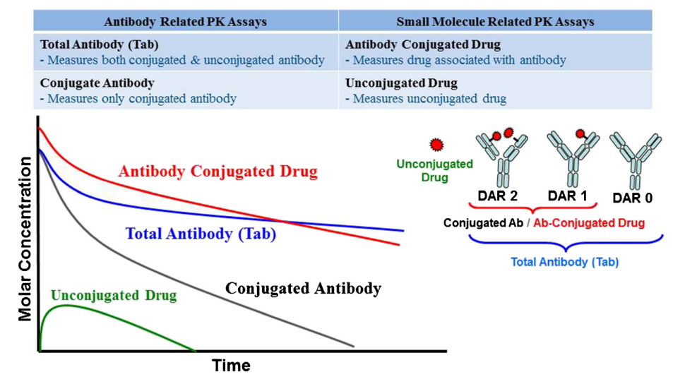
Figure 1. Pharmacokinetic profiles of different ADC analytes.4
The analytical strategies employed vary according to the physicochemical properties of the analyte. Ligand-binding assays (LBAs), such as enzyme-linked immunosorbent assays (ELISAs), remain the mainstay for quantifying large-molecule components such as total and conjugated antibodies. Conversely, small-molecule analytes-conjugated and free drugs often require advanced techniques like liquid chromatography-tandem mass spectrometry (LC-MS/MS), often incorporating sophisticated sample preparation protocols including antibody capture, enzymatic digestion, and selective drug extraction.4-5
Broadly, ADC bioanalytical methods can be categorized into two types: universal and specific. Universal methods employ generic reagents and standardized workflows to quantify common ADC components, offering applicability across various ADC platforms - an advantage for early-stage candidate screening and comparative analyses. In contrast, specific methods are precisely tailored to an individual ADC's unique molecular attributes, such as its linker, payload, or conjugation site.2 These methods provide superior specificity and mechanistic resolution but typically entail greater technical complexity and resource requirements.
This article provides a systemic comparative analysis of universal versus specific detection methods for ADC pharmacokinetic assessment. Using Trastuzumab-MMAE, HER2-targeting ADC, as a case study, we explore critical considerations that influence method selection, assay development, and validation processes. Our objective is to determine which analytical strategies yield optimal accuracy, sensitivity, and reproducibility for Trastuzumab-MMAE quantification, while establishing broadly applicable principles to guide rational PK assay development across ADC candidates.
Methods and Materials
Total antibody bioanalytical assay
Three methods were developed and evaluated for quantifying Trastuzumab-MMAE total antibody: a generic sandwich ELISA method, in which anti-human IgG polyclonal antibodies were both the capturing and detecting antibodies; a generic sandwich ELISA method, in which the capturing antibody was anti-human IgG monoclonal antibody, and the detecting antibody was anti-human IgG polyclonal antibody; an indirect ELISA method, in which the capturing reagent was human HER2 and the detecting antibody was anti-human IgG antibody Figure(2A-C).
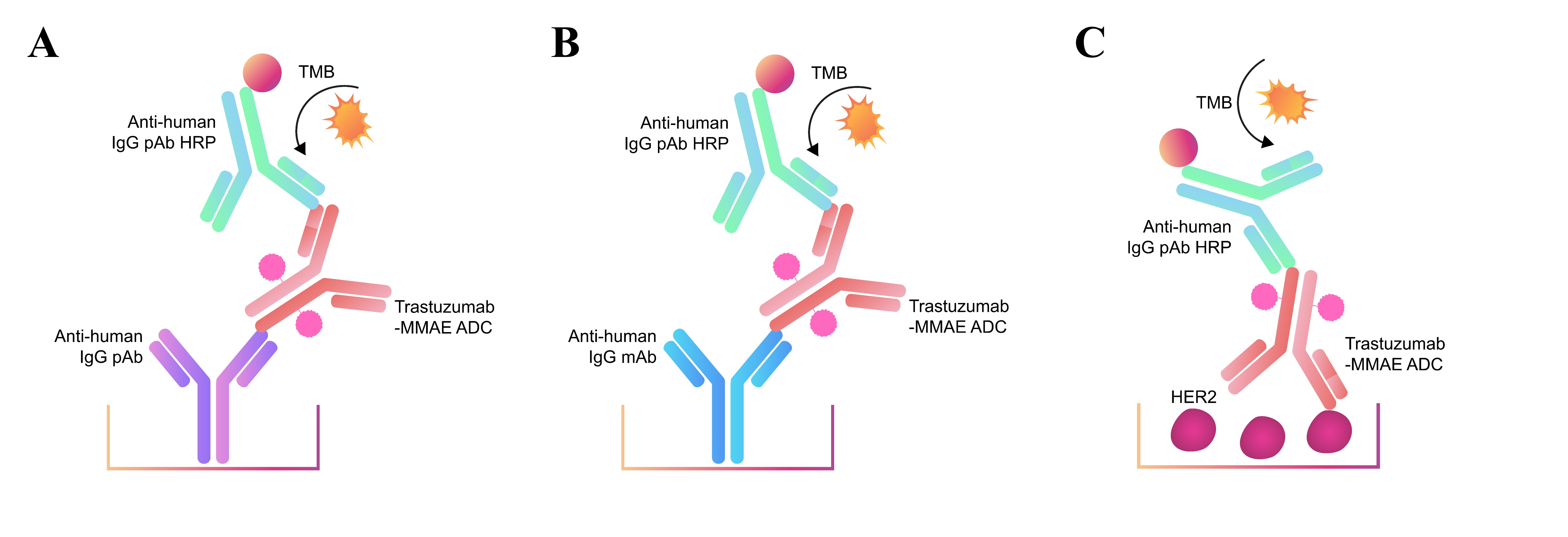
Figure 2. Three methods were developed and evaluated for quantifying Trastuzumab-MMAE total antibody
Specific ELISA for conjugated antibody
Two methods were developed and evaluated for quantifying Trastuzumab-MMAE specific antibody: a specific sandwich ELISA method, in which anti-MMAE antibody was the capturing antibody, biotinylated HER2 bound ADC and HRP-SA was the detecting antibody; a specific indirect ELISA method, in which anti-MMAE antibody was the capturing antibody and anti-human IgG monoclonal antibody was the detecting antibody Figure(3A-B). For the detection of ADCs, anti-payload Ab is critical for the assay development. Biotin-SA HRP can expand assay linearity range significantly and reduce the interference from serum.
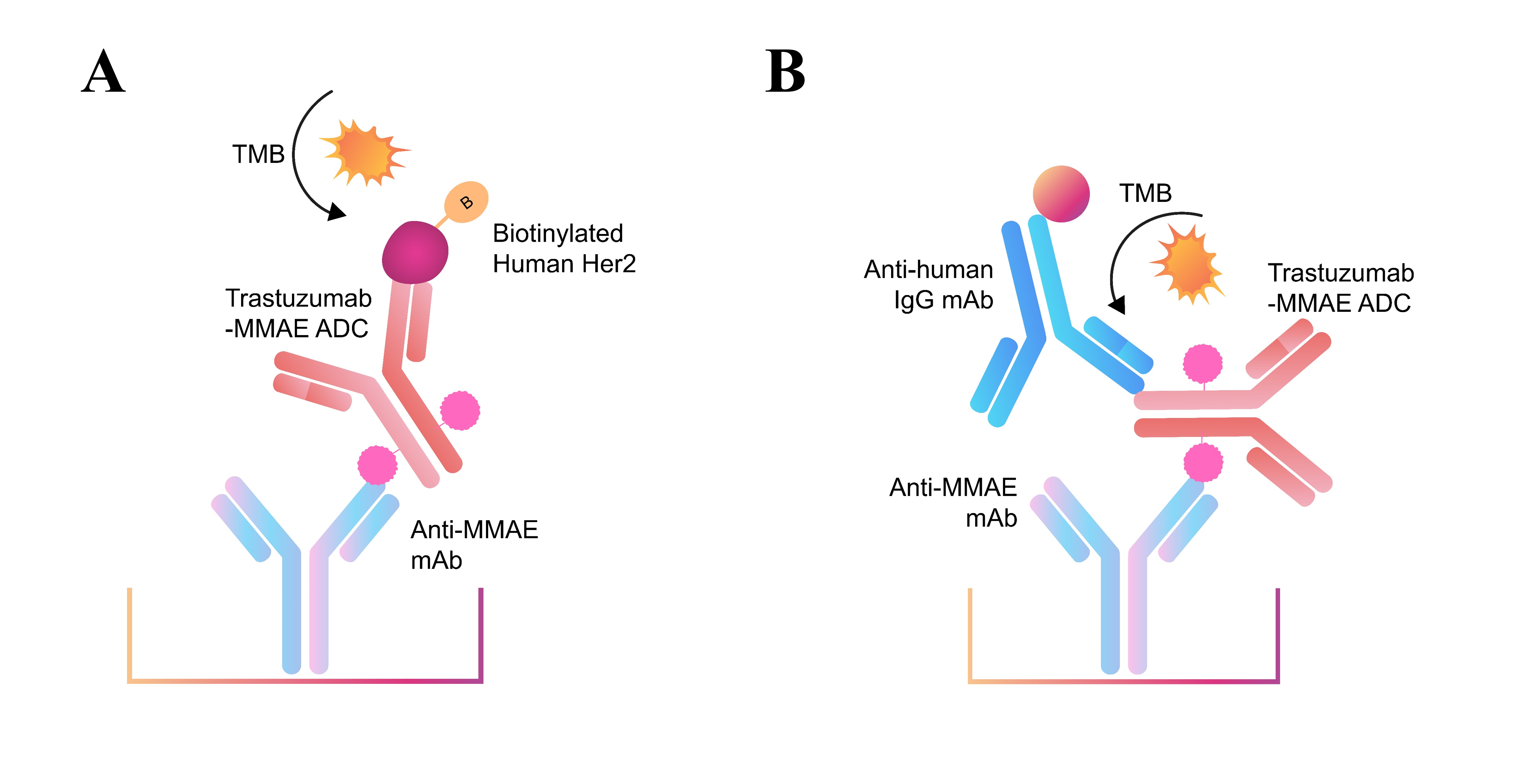
Figure 3. Two methods were developed and evaluated for quantifying Trastuzumab-MMAE specific antibody
Lyophilized proteins were reconstituted using ultrapure water into the reconstructed concentration and diluted as needed.
Table 1. Materials used for ELISA
| Materials | Catalog No. | Vendor |
|---|---|---|
| Affinity Purified Antibody to Human IgG (Fc) | 5210-0165 | KPL |
| Trastuzumab-MMAE | HY-164992 | MCE |
| Peroxidase AffiniPure Goat Anti-Human IgG, Fcγ fragment specific (min X Bov, Hrs, Ms Sr Prot) | 109-035-098 | Jackson |
| Monoclonal Anti-Human-IgG-Fc Antibody, Mouse IgG1 | IGG-S307 | ACROBiosystems |
| Monoclonal Anti-HER2 Antibody, Human IgG1 | HE2-M598 | ACROBiosystems |
| Human HER2, His Tag | HE2-H5225 | ACROBiosystems |
| HRP conjugated Anti-Human-IgG-Fc Antibody (6F11C8), mAb | IGG-LY69 | ACROBiosystems |
| Biotinylated Human HER2, His, Avitag, premium grade | HE2-H82E2 | ACROBiosystems |
| Costar 1 x 8 Stripwell, high binding EIA/RIA plate, flat bottom, without lid | 42592 | Corning |
| BSA | NA | Yancheng Saibao |
| TMB | A600954 | BBI Life sciences |
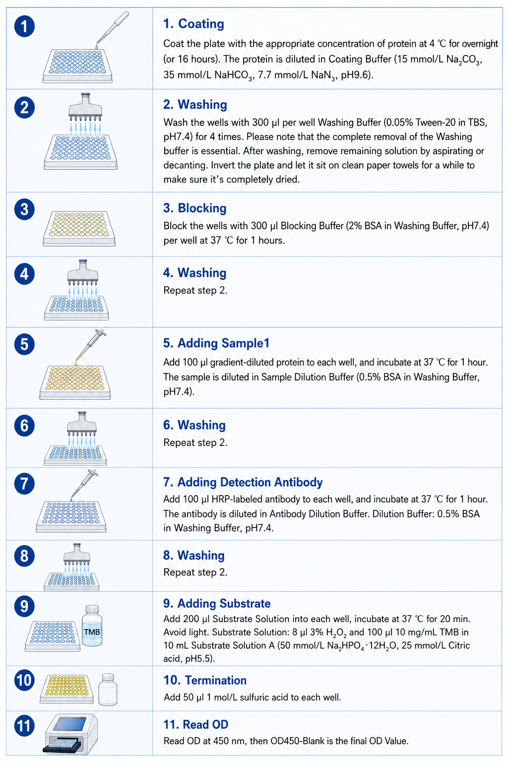
ELISA Protocol
Results and Discussion
Total antibody bioanalytical assay
We evaluated three distinct antibody assay formats for quantifying total antibody levels of ADCs: (1) a dual anti-human Fc polyclonal antibody (capture/detection) method, (2) a combination of anti-human Fc monoclonal (capture) and polyclonal (detection) antibodies, and (3) an indirect antigen-capture assay using anti-human Fc monoclonal antibodies. For Trastuzumab-MMAE, all three methods demonstrated excellent linearity across the concentration range tested and showed equivalent detection sensitivity for both unconjugated Trastuzumab and Trastuzumab-MMAE conjugate, confirming that drug conjugation did not alter antibody recognition (Figure4A-C). Comprehensive validation of the dual polyclonal antibody method (Method 1) confirmed superior performance characteristics with high standard curve accuracy (R² > 0.99), consistent inter-run precision (CV <15%), and recovery values between 90-110% of nominal concentrations, establishing it as the optimal approach for total antibody quantification in ADC pharmacokinetic studies (Figure 5A-C).
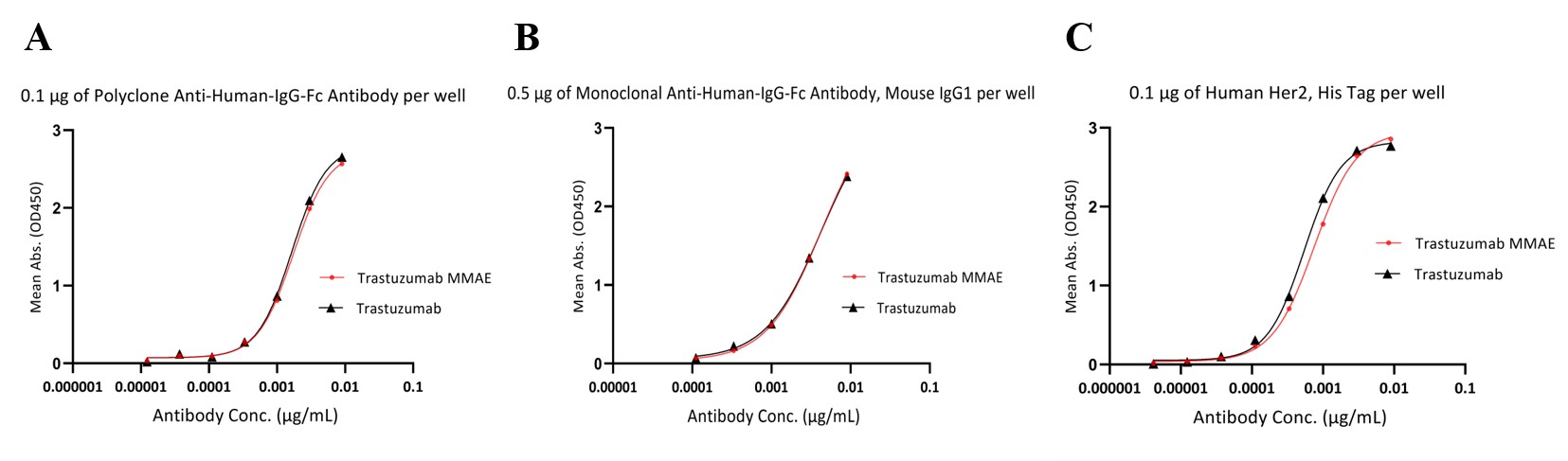
Figure 4. Comparative analysis of three methods the total antibody content of Trastuzumab-MMAE. A. a dual anti-human Fc polyclonal antibody (capture/detection). B. Hybrid method with anti-human Fc monoclonal (capture) and polyclonal (detection) antibodies. C. Indirect antigen-capture assay using anti-human Fc monoclonal antibodies.
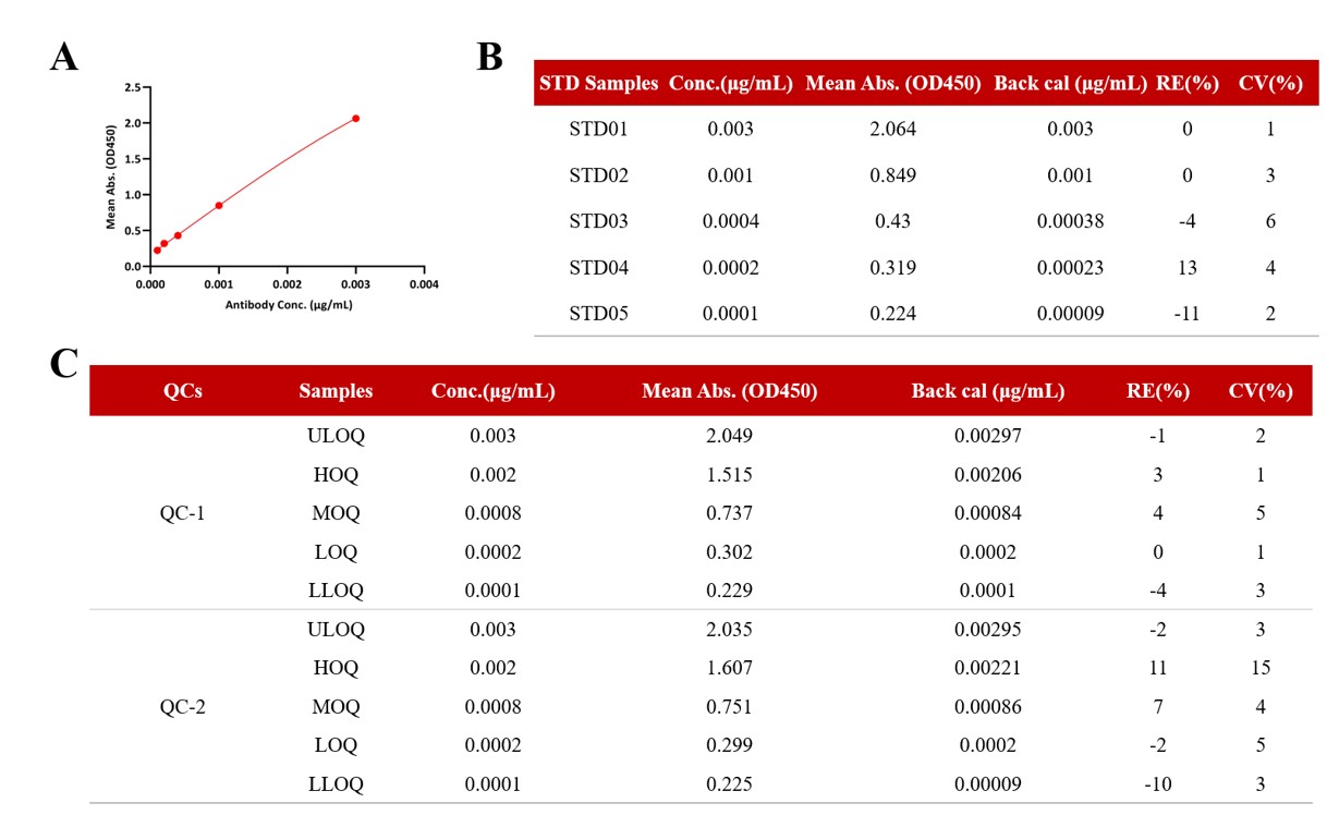
Figure 5. Method validation results for the selected dual anti-human Fc polyclonal antibody (capture/detection) method. A. Calibration curve fitting graph. B. Standard curve regression parameters and deviation assessment. C. Comprehensive accuracy and precision evaluation.
Conjugated antibody bioanalytical assay
We evaluated two methods for quantifying Trastuzumab-MMAE conjugated antibody. The first utilized a specific sandwich ELISA with anti-MMAE antibody as capture reagent, biotinylated HER2-bound ADC as bridging molecule, and HRP-conjugated streptavidin for detection. The second employed a specific indirect ELISA combining anti-MMAE antibody for capture and anti-human Fc monoclonal antibody for detection. Both methods demonstrated robust analytical performance with excellent linear ranges, and no significant differences were observed when using different anti-MMAE antibody clones, indicating method robustness (Figure 6A-B). Validation of the sandwich ELISA confirmed its suitability for precise conjugated antibody qualification, with a well-fitted standard curve (R²>0.99), inter-assay coefficients of variation below 15%, establishing this method as a reliable and reproducible for conjugated antibody quantification in ADC pharmacokinetic analysis (Figure 7A-C).
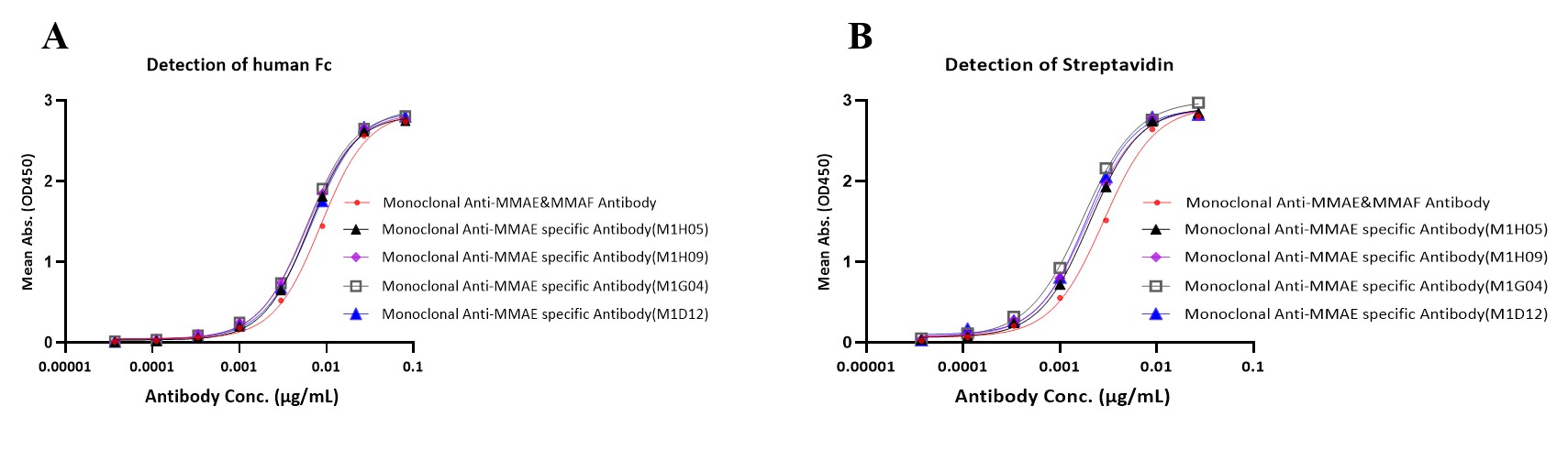
Figure 6. The results of two different quantitative Trastuzumab MMAE specific antibody. A. Detection of Fc. B. Detection of streptavidin.
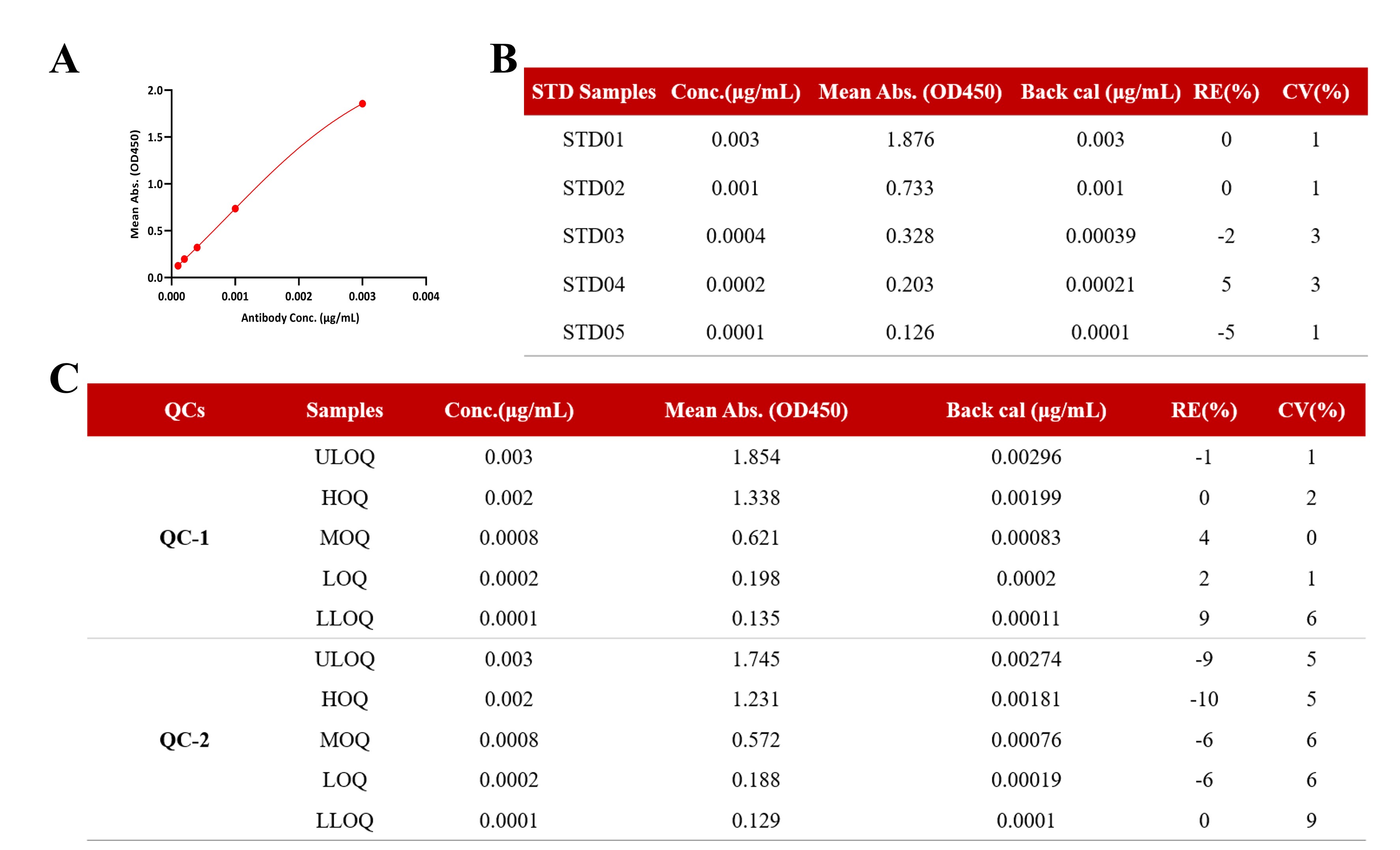
Figure 7. The methodology validation results of Detection of streptavidin. A. Calibration curve fitting graph. B. Standard curve regression and deviation. C. The results of accuracy and precision.
Conclusion
The ADC therapeutic landscape is experiencing unprecedented growth, with over 30 candidates advancing through clinical development pipelines alongside the 17 currently FDA-approved ADC. However, the relative scarcity of comprehensive pharmacokinetic data from approved ADCs means that bioanalytical strategies regulatory guidelines remain in continuous evolution. Our systematic comparison of multiple analytical methods for quantifying both total antibody and conjugated antibody fractions in Trastuzumab-MMAE demonstrates that well-optimized assays-whether employing universal or target-specific approaches-can achieve comparable analytical performance with respect to linearity, precision, and accuracy.
For practical implementation, we recommend selecting the optimal detection method through a holistic assessment of several critical parameters:
- Analytical flexibility across development stages
- Operational, complexity and resource requirements
- Resistance to potential matrix interferences
- Alignment with specific regulatory expectations for preclinical and clinical applications
The dual anti-human Fc polyclonal antibody method for total antibody quantification and the sandwich ELISA utilizing anti-MMAE antibodies for conjugated antibody determination represent particularly robust approaches that balance sensitivity with practical utility. ACROBiosystems offers rigorously validated anti-payload antibodies and complementary detection reagents that enable rapid implementation of these optimized methods, facilitating consistent pharmacokinetic assessment throughout of ADC development continuum from preclinical characterization to clinical evaluation.
Related Anti-Payload Antibody Products
>>> Click to learn more anti-payload antibodies
FAQ:
Q1: Which bioanalytical strategy is commonly used for ADC pharmacokinetic (PK) studies?
A: ADC pharmacokinetic studies typically require a multi-analyte strategy, including measurements of total antibody, conjugated antibody, conjugated drug, and free drug. Because each analyte reflects a different aspect of ADC disposition and biotransformation, no single assay can fully characterize ADC behavior in vivo. ELISA-based ligand-binding assays (LBAs) are widely used for total and conjugated antibody quantification, while LC-MS/MS is commonly applied to conjugated drug and free drug analysis. Together, these complementary approaches provide a comprehensive assessment of ADC exposure, stability, and payload release.
Q2: What is the difference between total antibody and conjugated antibody assays in ADC analysis?
A: A total antibody assay measures all antibody-containing species regardless of payload conjugation status (DAR ≥ 0), whereas a conjugated antibody assay specifically detects ADC molecules that retain one or more payload molecules (DAR ≥ 1). Total antibody measurements are commonly used to assess overall antibody exposure, while conjugated antibody data provide insights into payload retention, linker stability, and ADC deconjugation in vivo. Together, these assays offer complementary information for evaluating ADC pharmacokinetics and stability throughout development.
Q3: Why are anti-payload antibodies important for ADC bioanalytical assay development?
A: Anti-payload antibodies enable selective detection of payload-containing ADC species and are widely used in conjugated antibody assays. Unlike total antibody assays, they specifically quantify ADC molecules that remain conjugated to their cytotoxic payloads, making them valuable for evaluating linker stability, payload retention, and ADC integrity during pharmacokinetic studies. For payload classes such as MMAE, DM1, and DXd, antibody specificity and consistency can directly impact assay sensitivity and reproducibility. ACROBiosystems offers validated anti-payload antibodies and related reagents to support ADC bioanalytical assay development and PK studies.
Q4: What validation parameters are required for ADC ELISA assays?
A: ADC ELISA assays should be validated for accuracy, precision, sensitivity, selectivity, dilution linearity, recovery, and matrix effects. Depending on study requirements, analyte stability and lower limit of quantification (LLOQ) may also need to be evaluated. In addition to assay design, the quality and consistency of critical reagents can significantly influence validation outcomes and long-term assay performance. ACROBiosystems provides recombinant target proteins, Fc detection reagents, anti-payload antibodies, and other assay components commonly used in ADC bioanalytical workflows.
Q5: How should discrepancies between total antibody and conjugated antibody PK profiles be interpreted?
A: Differences between total antibody and conjugated antibody PK profiles can provide important insights into ADC stability in vivo. When conjugated antibody levels decline faster than total antibody concentrations, the data may indicate payload loss, linker cleavage, or ADC deconjugation. Accurate interpretation therefore requires reliable quantification of both analytes using appropriately designed assays. ACROBiosystems supports ADC PK studies with reagents and assay solutions for total antibody and conjugated antibody analysis, helping researchers evaluate ADC stability, payload retention, and pharmacokinetic behavior.
References
1. Fu, Z., Li, S., Han, S. et al. Antibody drug conjugate: the "biological missile" for targeted cancer therapy. Sig Transduct Target Ther 7, 93 (2022).
2. Mou S, Huang Y, Rosenbaum AI. ADME Considerations and Bioanalytical Strategies for Pharmacokinetic Assessments of Antibody-Drug Conjugates. Antibodies (Basel). 2018 Nov 30;7(4):41.
3. ProteoGenix. (n.d.). Assessing the pharmacokinetics of antibody-drug conjugates (ADCs).
4. https://www.proteogenix.science/scientific-corner/adc/pharmacokinetics-characterization/#importance-of-pkpd-analysis-for-adc-development
5. Qin Q, Gong L. Current Analytical Strategies for Antibody-Drug Conjugates in Biomatrices. Molecules. 2022 Sep 24;27(19):6299.
6. Mahmood, I. Clinical Pharmacology of Antibody-Drug Conjugates. Antibodies 2021, 10, 20.
 Popular ArticlesRelated RecommendationsPopular Events
 Dual-Payload ADCs: Translational Challenges and Clinical Pharmacology2026-06-16Page Views:413
 Next-Generation Dual-Payload ADCs: Target Selection, Payload Synergy, and Clinical Pipeline Analysis2026-06-12Page Views:2907
382-240.jpg) B7-H3 (CD276): Clinical Progress and Emerging Therapeutic Strategies in Solid Tumors2026-05-26Page Views:1123
B7-H3 (CD276): Clinical Progress and Emerging Therapeutic Strategies in Solid Tumors2026-05-26Page Views:1123 Beyond Expression: Functional Biology in ADC Target Evaluation2026-05-26Page Views:1026
 Navigating the Complex Landscape of ADC Quality Control: From Critical Quality Attributes (CQAs) to Clinical Success2026-03-19Page Views:2399




 Related Products